
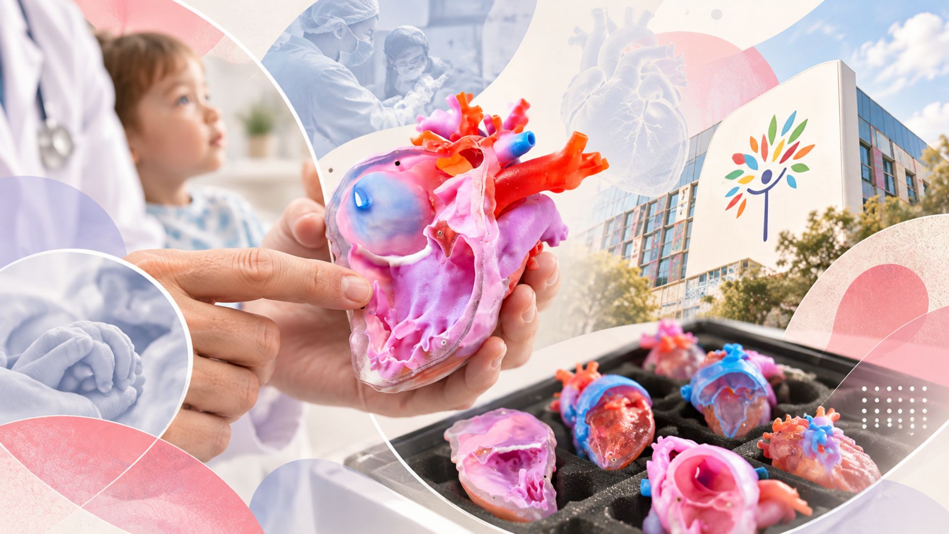
Small physical models of children’s hearts are helping clinicians explain congenital heart conditions to families at Melbourne’s Royal Children’s Hospital. A 2 October university feature describes Heartrium, a project that turns clinical scans into detailed objects people can hold, rotate and examine during a conversation about care.
The problem being addressed is one of understanding complex anatomy. A parent may be asked to make sense of chambers, vessels and a proposed operation while also processing difficult news. A flat drawing or an image on a screen can require considerable interpretation. A physical model gives the clinician and family something they can look at together, with the explanation tied to a visible structure.
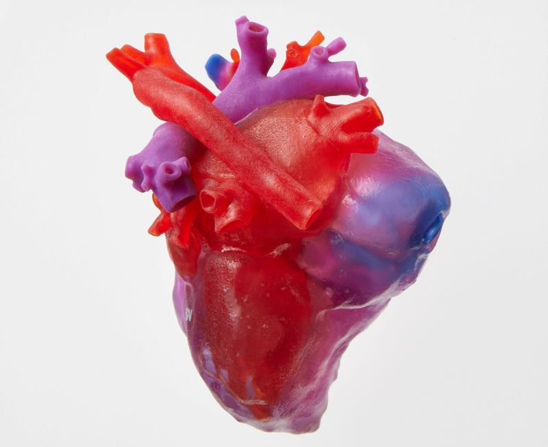
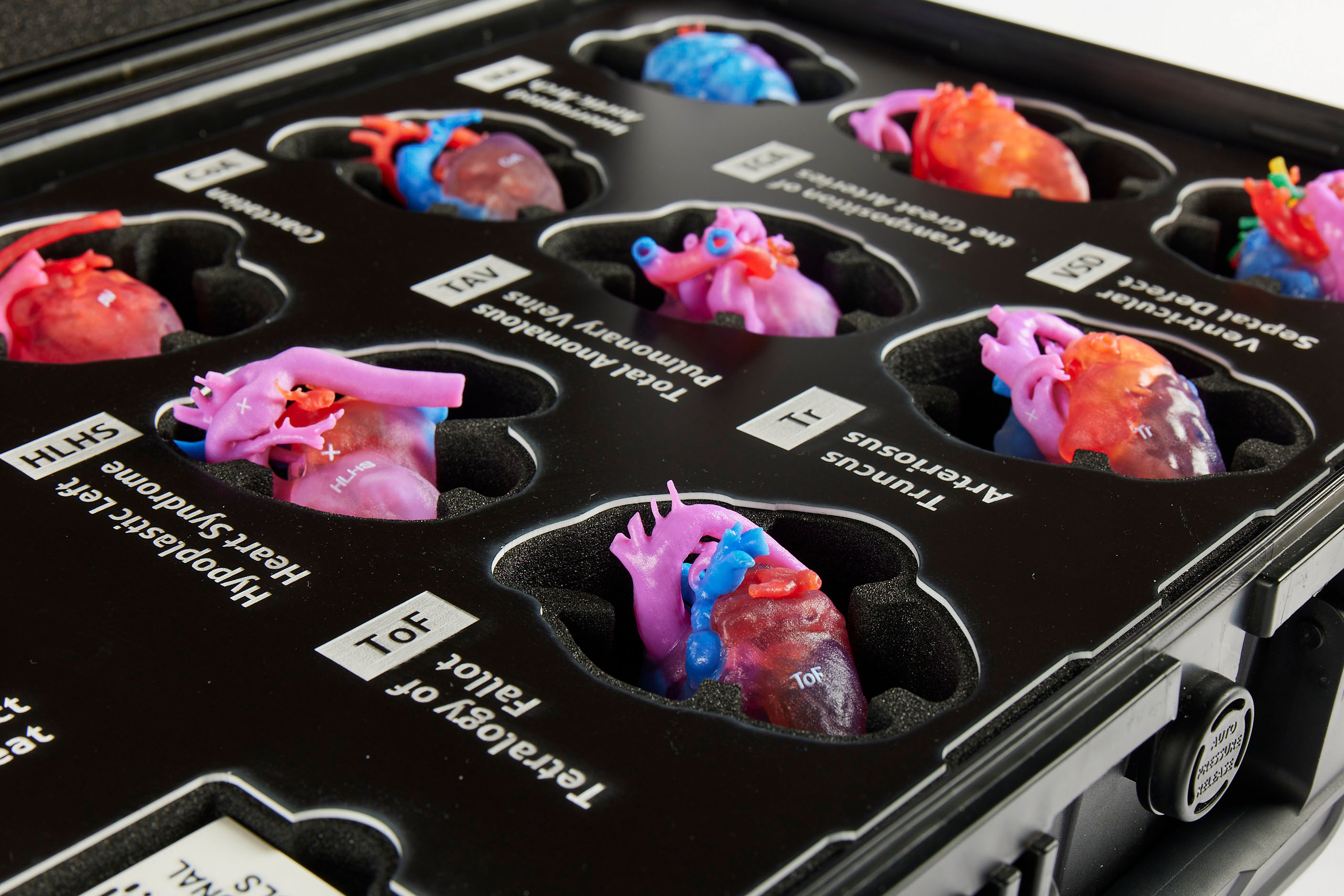
The project brings design and medical expertise into the same process. The project’s team description brings together Royal Children’s Hospital clinicians and Melbourne School of Design specialists in design, digital creation and fabrication. The university feature describes models used to explain conditions and surgical changes. It also recounts feedback from a family who found the scale and shape easier to understand when shown physically. That is a reported experience from the university’s coverage, not evidence that every family will respond in the same way.
Heartrium’s own model description explains how the objects are made useful for teaching. It says they are derived from echocardiography, CT and MRI imaging, reviewed by cardiologists and simplified with clinicians so that relevant features are clear. The life-sized models are soft and colour-coded, and are supplied in protective cases organised by condition. Clinical accuracy and understandable presentation both matter to that design task.
A model used to explain a condition also has limits. A standard example does not necessarily reproduce every detail of an individual child’s heart or every stage of their treatment. The clinician still needs to explain which features apply to the patient and which are illustrative. The university’s story itself describes a situation where an additional model was needed to represent a particular surgical stage, showing why a collection must develop around actual communication needs.
The latest coverage should not be confused with the project’s beginning. Heartrium’s project website says models have been evaluated with the Royal Children’s Hospital since mid-2024. Its separate digital models let users inspect internal structures and compare a condition with a normal heart on a phone, tablet or laptop. Those materials could help families revisit an explanation, while the physical kit remains available for consultations and staff education. Their advertised availability does not establish a measured clinical benefit on its own.
Evaluating the approach requires questions that go beyond how impressive the models look. Researchers could examine whether families understand the anatomy more accurately, retain information and feel able to ask informed questions. They would also need to consider language, accessibility and differences between clinical situations. Testimonials can identify useful experiences, but they do not substitute for a structured assessment of those outcomes.
For Australian hospitals, the wider lesson is about making information easier to discuss. Digital fabrication can turn scan data into a shared teaching object, but the model works within a conversation led by qualified clinicians. The new feature offers a concrete example of that collaboration in Melbourne. It does not establish better surgical outcomes or a newly launched treatment; it highlights an existing effort to help families understand the care being proposed.